Test Directory

TP53 mutation analysis

Containers - Adult

RedCapTubeEDTAKE2.7ml.jpg
Red Cap Tube EDTA KE 2.7ml
Volume Range

2 x 2.7ml peripheral blood or bone marrow specimen

Additive per Container

​EDTA

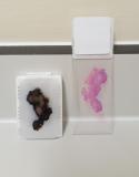
path_block_slide.jpg
FFPE block / H&E slide / Pathology report

Laboratory Site

RIE
51 Little France Crescent
Old Dalkeith Road
Edinburgh
EH16 4SA
Telephone: 0131 536 1000
WGH
Crewe Road South
Edinburgh
EH4 2XU
Telephone: 0131 537 1000

Transport arrangements

Fresh (peripheral blood / bone marrow aspirate) samples should ideally arrive within 24 hours of collection (maximum of 48 hours) and should be sent to the following address:


Western General Hospital
Haematology/Biochemistry Combined Reception
Immunophenotyping Laboratory
Crewe Road
Edinburgh
EH4 2XU


See also Specimen transportation.

FFPE specimens should sent directly to Molecular Pathology at the following address (see transport recommendations).

Molecular Pathology
Department of Laboratory Medicine
Royal Infirmary of Edinburgh
51 Little France Crescent
Old Dalkeith Road
Edinburgh
EH16 4SA

For patients with pathology specimens held within NHS Lothian there is no need to arrange transport of specimens.
If DNA has already been extracted from FFPE specimens, this may be sent to Molecular Pathology/HMDS at the WGH site.

Sample storage arrangements

Samples should be store at room temperature. See specimen requirements.

How to request

For Blood and Bone marrow samples: 

please refer to our detailed requesting instructions. The HMDS request form can be located here.

For FFPE samples: 

Testing for NHS Lothian patients can be requested by email to molecular.pathoogy@nhslothian.scot.nhs.uk.

Referral requests must be accompanied by a completed request form.

Please also refer to our detailed requesting instructions.


Availability

​Monday-Friday 9am-5pm

Anticipated turnaround

For Blood and bone marrow samples [CLL], results should be expected within 15 working days. See results.

For FFPE samples, an integrated Molecular Pathology report should be available within 10 working days. See results.

What happens if the result is positive or abnormal

​Requesting clinician will be contacted via telephone or email. Please ensure details are included on request form.

Static information/disclaimer

This test has been accredited to ISO15189. However, part of the procedure now makes use of an automated pipetting system which has been separately validated but is not currently accredited to ISO 15189.

General additional information

For assessment of TP53 loss (del17p) by FISH, please refer to the South East Scotland Cytogenetics Service.

CLL samples contain a mixture of leukaemia and non-leukaemia cells.  Since TP53 mutations are acquired within leukaemia cells, they will not be present within non-CLL cells within the sample. Therefore, it is important to be aware of the approximate percentage of leukaemia cells within the sample. The assay carries a limit of detection of 15% and therefore a low level TP53 mutation may not be detected by this approach.

For samples with a likely low level leukaemia percentage, Lymphoprep isolation of mononuclear cells will be performed. Hence, please inform the laboratory, where possible, of the total lymphocyte count and total white cell count in the sample

​TP53 mutation assessment is performed using Sanger sequencing to assess and identify sequence variants in exons 2a, 2d, 3, 4b, 5, 6, 7, 8, 10 and 11 of the TP53 gene (LRG_321 specific exon numbering). In most cases, a TP53 mutation is associated with loss of chromosome 17p [del17p]. However, some patients may carry a TP53 mutation without accompanying loss of chromosome 17p. Mutations in the TP53 gene are associated with chemotherapy resistance and poor prognosis in patients with CLL.

Assessment of the IGHV mutation status can also be performed.

For clinical advice on appropriate investigations and advice for the interpretation of test results, please contact us​.

Assessment of TP53 by Sanger sequencing is available for patients with endometrial carcinoma and to aid the diagnosis of high grade serous ovarian carcinoma. Where necessary, this may be supplemented by the use of next generation sequencing. Refer to Molecular Pathology Testing for further details